Open


October 29, 2026
The big toe provides one of the most important functions of our entire body when it comes to movement. In fact, the it carries the most weight of all the toes, bearing about 40 percent of our body weight. The big toe is also the last part of the foot to push off the ground before taking the next step.
With our reliance on our big toe comes two common foot conditions: hallux limitus and hallux rigidus. These two conditions are the medical terms for big toe arthritis. Specifically, they can be translated into the following from Latin: hallux = big toe; limitus = limitation; rigidus = rigid/stiff.
Big toe arthritis can be quite painful both because of the toe's weight-bearing responsibilities and the fact that we as humans are constantly on our feet. One giveaway sign of hallux rigidus is the presence of a bone spur on the top of the foot.
In this article, we break down the differences between the two halluxes, signs and symptoms, as well as non-surgical and surgical treatments.
Hallux Limitus: What Is It?
Let's start with hallux limitus since it's the earlier stage of hallux rigidus.
Hallux limitus is a condition in which the joint connecting your big toe to your foot (the first metatarsophalangeal joint - commonly called the first MTP joint) is inflamed, sore, and stiff. This condition is a form of degenerative arthritis (wear-and-tear of the cartilage within the joint) that occurs in the joint.
There are a few reasons why and how this condition develops:
Fallen arches: Fallen arches put excessive pressure on the big toe joint.
Excessive pronation: Excessive pronation puts excessive pressure on the big toe joint.
Overuse: Activities like running, doing lunges, or any excessive pressure to the toe joint can over time break down the joint.
Acute injury: stubbing or jamming your toe can damage the joint, and can worsen if you don't allow it adequate time to recover.
Hallux Rigidus: What Is It?
Hallux rigidus involves more progressive damage to the first metatarsophalangeal joint. This condition comes after hallux limitus. It means your joint has less range of motion, the cartilage is worn down, and there is likely the presence of osteophytes, also known as a bone spur. An osteophyte is a bony lump that grows around the joints.
In fact, hallux rigidus is the most common degenerative disorder of the foot.
So, what's the difference? Hallux rigidus is defined as pain due to an arthritic joint, whereas hallux limitus is defined as functional pain due to soft tissue tightness (i.e., when calf tightness limits the motion of the ankle joint) or a long and elevated first metatarsal bone (the bone behind the big toe).
These are the early signs and symptoms of hallux rigidus:
Pain and stiffness in the big toe during physical activity (walking, running, etc.).
Pain and stiffness aggravated by cold, damp weather.
Difficulty with certain activities (running, squatting).
Swelling and inflammation around the joint.
Loss of range of motion.
Depending on the severity of arthritis, the joint damage can be measured by the approximate range of motion of the big toe joint. Here is a breakdown:
Grade 0: 40° to 60° range of motion and/or 10% to 20% loss of range of motion.
Grade 1: 30° to 40° range of motion and/or 20% to 50% loss of range of motion.
Grade 2: 10° to 30° range of motion and/or 50% to 75% loss of range of motion.
Grade 3: ≤ 10° range of motion and/or 75% to 100% loss of range of motion.
Grade 4: Same as in grade 3 with more pain.
Does Hallux Rigidus Go Away?
Hallux rigidus is a progressive condition. It doesn't go away. However, there are a number of steps you can take (literally) to slow down the worsening of this toe condition. Treatment includes altering your footwear for shoes with firm supportive footbeds or rocker soles, avoiding activity that puts pressure on your big toe joint, and avoiding walking barefoot.
Is Walking Good For Hallux Rigidus?
Walking with the proper precautions and equipment should be OK with hallux rigidus. Walking in some cases, like with hallux limitus, can actually increase the range of motion. With hallux rigidus, the aim shouldn't be to increase range of motion but to reduce dorsiflexion (the action of raising your foot your toes upward). In this case, walking uphill can be painful. Walking barefoot can also be painful as there's nothing to protect the joint.
Do Orthotics Help Hallux Limitus and Hallux Rigidus?
Custom foot orthotics can provide multiple benefits for big toe arthritis. For hallux limitus, the goal of orthotics is to improve the range of motion within the first MTP joint. Conversely, for those with hallux rigidus, the goal of orthotics is to restrict the range of motion. Therefore, different orthotics are designed for different use cases and the severity of arthritis. As those with fallen arches or excessive pronation are particularly susceptible to developing hallux rigidus, custom orthotics to correct these biomechanical abnormalities can also help.
One such approach is using Morton's extension or a reverse Morton's extension on a supportive orthotic. This approach utilizes a stiff orthotic material that extends under the big toe from the base to the tip. The goal here is to shift weight off of the weight-bearing joint of the big toe.
Additional measures beyond orthotics include shoe modifications. Shoes should be sufficiently long, comfortable, with a high and broad toe box, and should have enough space for the orthotic device. Additionally, shoes with a full-length carbon plate or built-in rocker sole can reduce pressure on the big toe, and should be explored as an option.
How Successful is Surgery For Hallux Limitus and Hallux Rigidus?
There are two primary surgical procedures to treat big toe arthritis: a cheilectomy and a first MTP joint fusion. A newer surgical procedure is emerging as well, in which an artificial joint (Cartiva) is inserted to replace the damaged joint. Orthopedic surgeons perform these operations. In general, non-surgical procedures should be explored and exhausted before proceeding to surgery. But, in some advanced cases, patients can greatly benefit.
Cheilectomy
A cheilectomy is a surgical procedure to remove excess bone from the joint of your big toe. It's used for mild-to-moderate cases of hallux rigidus. The intention is to decompress the joint area by removing the excess volume of bone. The most common form of a cheilectomy is to create a keyhole incision at the top of the foot, where a tool is inserted to remove the bone spur. This procedure is typically done under local and general anesthesia. Although the range of motion won't necessarily be improved, the removal of the bump can help reduce pain and discomfort.
The procedure typically takes less than 30 minutes and patients can be in and out of the surgical facility within 2-3 hours. Initial recovery includes two weeks in a soft cast and wearing an orthopedic sandal with light weight-bearing activity. The cast and stitches are removed roughly two weeks post-op. Long-term recovery includes returning to activity within 4-6 weeks although swelling and pain can resume for up to six months.
First MTP joint fusion
In a first MTP joint fusion, the bones are joined (fused) together permanently so they cannot rub against each other and cause pain. This operation is typically performed for moderate-to-serious cases of hallux rigidus when osteoarthritis has severely damaged the first metatarsophalangeal (MTP) joint.
Similar to a cheilectomy, an incision is made on the top of the big toe, and cartilage is cleared away to allow the two bones to heal together. For this operation, the two bones are fused together using screws and/or plates. This permanent procedure reduces all range of motion but eliminates the pain-causing joint.
Post-op, you'll be examined at the two-week, six-week, three-month, and six-month intervals. X-rays may be taken as well to assess the growth of the fused bones. You can expect recovery to last up to six months.

August 15, 2024
Whether they're discoloured, cracked, ingrown, curly, or on the verge of falling off, toenail anatomy can be fragile and our nails can do funny things. A less commonly discussed issue is a secondary toenail. Also known as an accessory toenail or petaloid toenail, a secondary toenail is a sneaky little extra nail twin that grows alongside or beneath the pinky toenail. This abnormal toenail growth can be concerning, so it’s natural to have questions.
Signs You Have a Secondary Toenail
It’s important to know the difference between an ingrown toenail and an accessory toenail.
First, ingrown toenails can grow on any toe, whereas a secondary toenail appears on the fifth, smallest toe.
Ingrown toenails are also far more common, and pierce the skin rather than growing alongside the nail.
Secondary toenails are thin in nature and often don’t cause pain, infection or swelling like ingrown toenails do (although it is possible).
Lastly, ingrown toenails form on the side of the nail unit, whereas secondary toenails grow underneath the proper nail.
What Causes a Secondary Toenail?
A secondary toenail is a rare phenomenon.
It often makes an unexpected appearance after trauma or injury to the nail bed. You may notice your nail growing differently after stubbing or hurting it. Chronic pressure from tight shoes or less-than-perfect nail trimming can also coax this extra nail into existence.
Do You Need Surgery For Secondary Toenails?
Studies show that the treatment of choice for this extra pinky toenail is an excision, especially if it’s infected. A partial or total nail avulsion from Toronto’s Feet First Clinic may be the solution you need, but you’ll have to schedule a proper foot assessment to determine the best course of action.
Treatment of accessory toenails targets the original toenail, not the second one. The second toenail should grow properly after the “main” nail is removed.
March 9, 2023
Plantar warts and sports are an "iconic" duo of foot conditions and risk factors. Many athletes are looking for a quick fix for their plantar wart woes. But the truth is, switching your mindset towards prevention and medical foot care is just as important.
Let's look at our plantar wart guide for athletes and gym-goers! We'll answer some of your most pressing questions and dive into the following:
What are plantar warts?
Why are athletes prone to plantar warts?
Preventing plantar warts: foot hygiene and foot care
Prevention plantar warts: shoe choices
What are Plantar Warts?
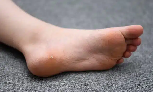
Plantar warts are viral skin infections caused by the Human Papilloma Virus(HPV) that appear on the bottom of the feet.
Many people assume that HPV is solely a sexually transmitted infection, but there are over 200 strains, some causing warts and others causing cancer. People usually contract this virus strain by walking or running barefoot on an infected surface, which enters the skin via contact or through small cracks in the skin.
Warts appear round and look deceptively like corns, calluses and blisters. They result from the body's failure to fight off the HPV, which can sometimes disappear on its own depending on the strain. Symptoms and signs of plantar warts include:
A hard and "grainy" growth on the bottom of the foot, often on the ball of the foot or base of the toes
On dark skin, it may appear lighter than the surrounding skin
Calluses surrounding the wart if it has started to grow inward
"Wart seeds" - which look like a small black dot in the wart
Pain when walking or touching the wart
Mosaic warts or a cluster of warts
Treatment options from a foot clinic include cryotherapy and needling. You can read about treatments in more detail here.
Why are Athletes Prone to Plantar Warts?
Plantar warts have the potential to debilitate athletes and gym-goers and severely impact their physical progress. But why are the physically fit more likely to contract them in the first place?
HPV loves warm, moist environments. These include communal showers and locker rooms where sports teams typically congregate without shoes or socks. So without adequate foot protection, members of the same team can unknowingly pass on this troublesome skin infection to each other.
Competitive swimmers are also at risk since the walking area surrounding the swimming pools is also communal with heavy, barefoot traffic. Furthermore, the rough surfaces of pool decks may cause micro-trauma to the bottom of the feet, increasing the risk of viral contraction.
Athletes and fitness lovers are also more likely to experience hyperhidrosis, or excessive sweating of the feet, which can increase the risk. Athletes often also share infected gym equipment, such as mats, without thorough cleaning.
Photo by Anna Shvets from Pexels
Plantar Wart Prevention: Foot Hygiene and Foot Care
The best way to prevent plantar warts is to strictly monitor your foot health and keep your feet clean. If you already have plantar warts, these practices can help prevent the condition from worsening.
Here are some practices to abide by:
Follow a daily foot care routine, being careful not to miss days when you play sports or go to the gym. It should include cleaning, exfoliation, moisturizing, and more. Here is an excellent one to follow!
Lightly dust your feet with baby powder. This can also help with foot odour! You can also try Gehwol Foot Powder.
Wear moisture-wicking socks, foot pads or other protective products.
Never go barefoot in a communal area like a locker room, shower or pool deck. Always wear protective footwear like sandals or shower shoes while in these spaces.
Don't scratch or pick at any pre-existing plantar warts.
Air out your shoes after using them.
Always wash your hands if you touch one of your plantar warts.
Don't share shoes, socks, towels and other products with other people.
Plantar Wart Prevention: Shoe Choices
Athletes can't avoid keeping their feet cooped up in runners all day. What they can do, however, is make shoe choices that help to prevent the growth of plantar warts.
Shoes should be breathable and waterproof. You know how some runners have those mini holes in the upper that let air enter and escape? Those are the ones you should be on the hunt for! The ventilation keeps your foot environment fresh and healthy while you spend the day exerting energy. When shopping for shoes or attending a shoe fitting, stress the importance of finding shoes like these by asking for breathable options.
You can also look for shoes made with GORE-TEX fabric, which repels moisture while letting water vapour from sweat exit the shoe.
Looking For More Info on Plantar Warts?
We know that athletes are at risk of developing plantar warts, but did you know children are also vulnerable? If you're a concerned parent, check out this blog on managing plantar warts in children.
September 15, 2022
Warts can occur anywhere on your body. These small, circular growths are a type of skin infection that stand out because of their roughness, slight discolouration, and sometimes even pain. When it comes to your feet, the soles are a common place for warts. This skin condition is known as plantar warts.
Although foot warts are a common occurrence, this condition comes with very treatable solutions. From self-care remedies to visiting a foot specialist, the scale of treatment methods varies based on the persistence level of warts. If you're looking to avoid plantar warts altogether, learn about what causes them, or how to treat them, you've come to the right place.
In this article, we explore plantar warts, their causes, how to treat foot warts, and finally how to book an appointment with a foot specialist if you have persistent plantar warts.
What are foot warts?
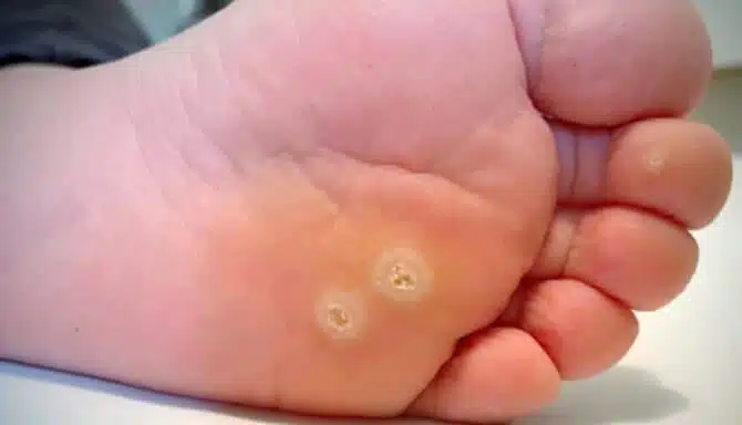
Plantar warts are common viral skin infections that can grow on any place on your foot. Usually the most common area is the bottom of the feet. They start out small, flat and look like a corn, callus or blister. Walking puts pressure on the wart causing a hard layer of skin to build up overtop. Applying pressure to plantar warts (walking, standing, or running) may be uncomfortable, if not painful.
Anyone can get plantar warts. They are not a severe health condition and sometimes go away on their own. However, they are contagious. If they persist, they can grow, spread and cause foot pain when you stand or walk on them.
What causes plantar warts?
Human Papilloma Virus (HPV) causes foot warts by infecting the foot's outer layer of skin. HPVs are a group of more than 200 related viruses. In the case of plantar warts, the strain of HPV causing this foot condition is not severe and varies from the strain that infects the reproductive tract. So, if you think plantar warts and herpes or genital warts are related, don't worry - they're not.
The virus can enter the foot by:
A cut or perforation in the skin
If the skin is too dry
If the skin is too wet
If you come into contact with a contaminated surface: Frequenting high-traffic common areas like showers, change rooms, and pool decks increase your likelihood of contracting the HPV that causes warts because the virus spreads more easily in these areas. Wearing sandals or flip-flops will help lessen your chances of catching warts by ensuring your bare feet do not come in contact with the virus that causes them.
Children can be more prone to foot warts in part due to having sweaty feet
As warts are caused by a virus, if you have a weakened immune system then you can be more susceptible to foot warts
Why Are Foot Warts Difficult To Treat?
The wart goes into your body's skin cells. It uses those cells to manufacture more wart tissue. Your body doesn't recognise it as foreign invador, as the wart is within your cells. As it grows the foot wart takes up nerves and small blood vessels. You might see black dots within a foot wart. These are called petechiae and are small burst blood vessels - not dirt.
So, what happens when you get plantar warts? Fortunately, there are several treatment options, including DIY methods as well as visiting a foot specialist.
But, please note: If you have low immunity, are prone to infection or have conditions that discourages "DIY treatments" (like Diabetes and Rheumatoid Arthritis) then you need to seek professional help to treat the warts.
It is also worthy to note that regardless of treatment, warts can come back. They are stubborn and usually multiple treatments are required to get the foot warts under control, especially if they are large.
8 Foot Wart Prevention Tips
Regardless of the treatment method you choose, it's best to follow these practices:
Cover the wart to help prevent it from spreading to either other people or another part of your body
Wash your hands any time you touch the wart - accidentally or intentionally
Do not pick the wart; you can transport it to your fingers
Make sure other members of your household do not have foot warts
Wash down the bath/ shower after each use
Figure out why you caught the wart in the first place. If it's a weekly swim, then wear footwear around the pool and in the communal showers. If your feet sweat, then try and reduce those sweating factors.
If you are using over the counter treatments then gently dry buff the hard skin of the foot wart. This allows treatment to be more effective. However you should not use that file anywhere else.
Replace the insole of your commonly used footwear so any wart tissue within your shoes is not going to reinfect you. If the insole is not removable, spray the inside of the shoe with a disinfectant spray that kills viruses (like Lysol disinfectant spray).
Sometimes (and rarely), plantar warts will get recognised by the body. It may take some years but they can go away by themselves. If that's not the case, a foot specialist like a chiropodist can easily (and more quickly) remove plantar warts. There are also different treatments depending on how resistant the wart is to certain methods. For instance, if the wart is not responding to one treatment, a more aggressive treatment may be tried.
Below you'll find a mix of at-home treatment methods and procedures done by a foot specialist at a local clinic:
Plantar warts treatments
The most effective ways to treat foot warts are classed as either chemical or non chemical treatments.
Chemical
Blister-Inducing Medicine
View this post on Instagram
A post shared by Feet First Clinic (@feetfirstclinic_)
A foot specialist can help induce a blister under the wart by applying cantharidin, a type of burn agent. This is applied every 2 to 3 weeks by a chiropodist. The cantharidin forms a blister over the wart, which separates the wart from the skin covering it and brings the wart to the surface. The blister then dries over the next few days and the wart falls off. The treatment is repeated until the wart is fully removed. Although the blister can be painful for a few days after treatment, cantharidin is considered more effective than other, less aggressive treatments - especially if you have stubborn persistent warts that don't respond to more conservative treatments.
Cryotherapy
Cryotherapy is a method by which extreme cold is used to freeze and remove a plantar wart. Depending on the freezing solution, it can affect just the top layers of the wart or it can go down deep. Over the counter freezing solutions are not too strong and typically are only effective for very small and newly formed warts. Strong freezing comes in the form of liquid Nitrogen and is usually done in a clinical setting. It can be sore as it touches some of the nerves within the wart - it is also extremely cold. One of the main side effects is frozen blistering. Usually it goes in a day or two, but if it is sore or the blister persists then you should see your foot specialist.
Like cantharidin, cryotherapy requires repeat treatments every 2-3 weeks in order to be effective. It is not as aggressive as cantharidin, and therefore the wart typically takes longer to go away.
Salicylic Acid
Salicylic acid is a topical medication that is applied to the wart. It softens the skin and helps shed layers of skin (and the wart) bit by bit. It is found in many over-the-counter wart removal products, and a chiropodist can prescribe a high-strength salicylic acid topical solution that you can apply regularly at home. The higher the dose of salicylic acid, the better its treatment. You will see it working as the skin will turn white. Sometimes there is a slow build up, and then it rapidly affects the wart. While Salicylic Acid may be less painful than cyrotherapy or cantharidin, it takes much longer to treat the wart.
Salicylic acid can be painful if not properly washed off or managed properly. Your chiropodist will tell you how to properly manage this treatment.
Non-Chemical
Laser Treatment
Laser treatment involves cauterizing blood vessels. Ouch, that sounds intense. But, your medical practitioner will numb your skin first. To be effective, this treatment should be repeated every 2-4 weeks.
Needling
Needling is a simple, but more invasive wart removal procedure with roughly 70% effectiveness. After a local anesthetic is administered, a specialist punctures the wart with a sterile needle about 100 times. This method pushes the HPV away from the surface of the skin and further into your body, where an immune reaction is triggered. If successful, your body's immune system will do the rest.
View this post on Instagram
A post shared by Feet First Clinic (@feetfirstclinic_)
Excision
Excision is used for plantar warts that resist other treatments. The foot is first numbed with a local anesthetic. Then, as the name implies, the wart is cut away (or “excised”) from the skin. The base is also cauterized to ensure no living viral tissue is left behind. There is a risk of scarring, so this method isn’t used unless other treatments have failed.
Other "treatments" for foot warts you may have heard about
Duct tape: Duct tape is a common yet relatively unproven method for treating plantar warts (largely because plantar warts can disappear on their own, which makes it difficult to attribute to the treatment method). This DIY method relies on constant use of duct tape and allowing the wart to get so wet and damp that it goes away. Very few people have had success because keeping the duct tape on for such a long time is very difficult. It is also not too hygienic; you'll dampen the surrounding skin, which can cause other issues.
Banana skins: There are supposedly enzymes within the actual skin of the banana that helps with wart treatments. Again, the success rate is extremely low.
Apple Cider Vinegar: Success rate of this is extremely low.
Salt water bathing: This doesn't do anything apart from dry your feet out, which will actually make you a more prone to foot warts
Rubbing a coin on the area: This method has been mentioned multiple times in internet forums; supposedly, the wart goes away in 1 day with this method. However, we've never seen this work and there is no scientific evidence that it does. For those that claim it does work, then it's highly likely it wasn't a wart in the first place.
Not only are the above "treatments" simply ineffective myths, they can waste precious time while your wart grows, spreads and multiples. At the end of the day, the best way to treat plantar warts is to see a foot specialist like a chiropodist at the first sign of any symptoms. The earlier you start treatment, the quicker and more effective it will be.
May 9, 2022
“Broken foot” is a broad and general term. Which bone did you break exactly? Each foot contains 26 bones and 100+ joints, muscles, tendons, and ligaments making it a complex network of parts.
A broken bone is the most serious bone injury. It involves the partial or complete breaking of a bone. It can be either a crack or a break.
Think of your bones like an HB pencil. It can bend. But there's a breaking point when you exert too much pressure. Although bones are incredibly resilient, once you go beyond a bone's breaking point, it cracks or fractures.
In this article, you'll learn about the types of bone breaks, common broken foot bones, recovery time, and ways to diagnose the severity of the injury.
Types of bone breaks
There are several types of bone breaks, as well as two categories of the displacement of broken bones.
Stress fracture: A stress fracture, or hairline fracture, is a crack in the bone. This is often caused by repetitive movements that over time cause the bone to weaken and ultimately crack. This is the least severe form of a broken bone and is common in the leg and foot, namely the tibia, femur, and metatarsals.
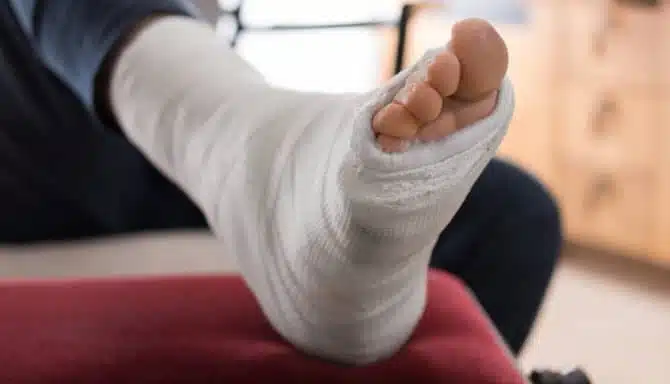
Stable fracture: A stable fracture is a clean break in the bone where the bone maintains its alignment. This means that no surgery is necessary. Instead, a cast may be sufficient to stabilize the bone while it heals.
Open (compound) fracture: A compound fracture is when a bone breaks and pierces through your skin. This severe injury often requires surgery, both to close the skin to prevent infection, and to re-align the broken bone.
Transverse fracture: A transverse fracture is an injury where the bone breaks at a right angle (i.e.: in a straight line perpendicular to the length of the bone). This type of injury on an X-ray would be analogous to sawing the trunk of a tree.
Oblique fracture: Whereas a transverse fracture is straight across bone, an oblique fracture is a broken bone that occurs at an angle. On an X-ray, this would look like a bone with two pointed edges close to or at the breaking point.
Spiral fracture: Also called a “torsion fracture”, a spiral fracture involves a twisting force. Like an oblique fracture, the bone breaks at an angle; however, the extreme twisting force also causes a component of the break to be parallel to the length of the bone.
Comminuted fracture: A comminuted fracture is when a bone breaks in multiple locations. This severe injury is usually the result of a major accident, like a crash or bad fall.
Avulsion fracture: An avulsion fracture involves the detachment of a bone (or piece of the bone) from the tendons and ligaments it’s attached to. Avulsion fractures are most common in the ankle.
Not only are there different types of fractures, but there are also two categories for the extent of the break. These are non-displaced fractures, in which broken bones generally maintain their original alignment, and displaced fractures, where a broken bone requires re-alignment because it's forced out of position.
Most common broken foot bones
Metatarsals: Your metatarsals are the small but mighty bones in your toes. Each of your toes has a metatarsal and are labeled from one through five, starting with your first (big toe). The fifth metatarsal (aka your pinky toe) is prone to breaks because of the regularity of stubbed toes, whether it's in your home, at work, or on the field. (Read more on what to do when/if you stub your toe.) That's not to say your other metatarsals aren’t also prone to breaks. Each of your toes can break, especially if you're someone who lifts and drops items or works in a labour-intensive industry.
Tibia: Your tibia, also known as the shin bone, is prone to breaks, both from one-time trauma or repetitive usage. As a weight-bearing bone, this is a common site of stress fractures. It's also prone to full breaks as it's the connector between your ankle and knee. This is a commonly broken bone in sports where your lower legs are targeted, like basketball, football, hockey, or soccer.
Heel bone: Your heel can break from falling and landing on your feet. For example, you may fall down the stairs feet first and injure your heel, or you could fall off a ladder. Other kinds of trauma impacting the foot, like a car accident, can also break your heel.
Causes of Broken Foot Bones
The most common causes of fractures are:
Trauma: Trauma refers to an acute injury to the bone causing it to break. This is usually a one-time event that forces a bone beyond its breaking point. Common scenarios include falls, car crashes, collisions during sports, or even stubbing your toe.
Osteoporosis: Osteoporosis is a disease that weakens the bone. Specifically, it occurs when bone mineral density is reduced, making breaks and fractures more common. This condition is common in women after menopause (the 12-month timeframe after a woman's last period) but it can also occur in men and premenopausal women. Hormonal disorders, chronic diseases, and smoking can all contribute to reduced bone mineral density, lack of calcium or other mineral deficiencies in bone composition.
Overuse/stress: Minor, but repetitive trauma to a bone can cause it to crack and break. These repeated forces over time don't necessarily push your bone beyond its breaking point at once. Rather, the consistent impact causes slow degradation and weakens a part of your bone to the point where it can no longer recover between uses. These overuse injuries are commonly referred to as stress fractures. A precursor to a stress fracture is a stress reaction, which is a deep bone bruise. Stress reactions can turn into a stress fracture if not treated. Common overuse injuries occur in long-distance runners, soccer players, and jumping up and down repeatedly.
How long does it take for a broken foot to heal?
There's no exact timeline but bones can take anywhere from 6-12 weeks to heal. Recovery largely depends on the severity of the fracture, as well as your recovery efforts. Additionally, whether the broken bone is weight-bearing or not has an impact on recovery time. Generally, children and young adults heal faster, whereas the older population will have delayed recovery.
Your diet and eating habits also influence recovery time. A diet rich in calcium and vitamin D helps restore bone tissue (vitamin D helps your body absorb the calcium). Foods rich with calcium include dairy products like milk, cheese, and yogurt, as well as non-dairy foods such as almond milk, orange juice, cereals, broccoli, greens, beans, and fish. Foods with plenty of Vitamin D include mushrooms, soy milk, oatmeal, fish, and eggs (especially the yolks!).
How can you tell if you break a bone?
The fastest and most reliable method to diagnose a broken bone is for a doctor to perform medical imaging. X-rays are a common method to diagnose fractures and broken bones, whereas MRIs are done to examine muscles, tendons, and ligaments.
However, there are other symptoms and signs of a broken bone. These include, but are not limited to:
Hearing a snap: Except perhaps in the case of ankle popping and cracking, a snapping noise is indicative of a broken bone.
Pierced skin: As bad as it sounds, a more obvious way to tell if you have a broken bone is if it's visible, namely in the case of compound fractures.
Location: Where it hurts can tell you a lot about whether you've broken a bone or not. Pain localized to the bone itself is a good indicator compared to, say, pain in soft tissue, in which case it may be a muscle or tendon injury rather than a bone injury. If the bone is painful to touch, it may be a broken bone.
Unable to bear weight on it: With broken bones, especially weight-bearing bones, putting any sort of weight on it is painful. That said, broken bones, depending on the severity and time since injury, may be painful when you have little to no weight on the joint.
Swelling: Common with a broken foot is bruising and/or discoloration. This association is however challenging. Swelling is a sign of a number of conditions and injuries unrelated to a broken bone.
If you experience any of the above signs and symptoms, you should immediately seek medical attention and get medical imaging.
November 2, 2021
What's that bump on the top of your foot? It could be a bone spur. It could be a blister. It could even be a wart, or a corn. It could also be what's known as a ganglion cyst. Like a bone spur and a blister, this is a visible condition that commonly affects the top of the foot, as well as the ankle and heel. They can also occur in other areas.
In this article, you'll learn more about ganglion cysts, how to spot them, treatment methods, and the severity of this foot condition. Additionally, we'll outline some distinctive features so it's easier to differentiate between a ganglion cyst and other common foot conditions.
But first, let's start with the first burning question: what is it?
Ganglion cyst: What is it?
Ganglion cysts are noncancerous lumps - or nodules - that commonly develop along the tendons or joints of your wrists or hands. Specifically, these are the most common spots:
The base of the fingers on the palm, where they appear as small pea-sized bumps
The fingertip, just below the cuticle, where they are called myxoid cysts
The outside of the knee and ankle
The top of the foot
As foot specialists, we're going to focus on ganglion cysts on the foot - namely the top of the foot and along the ankle.
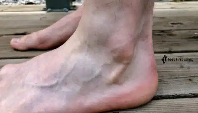
In terms of what this condition looks like, ganglion cysts are often round or oval in shape. They're filled with a jelly-like fluid. They're quite small - roughly the size of a pea - but can grow to roughly 2.5 cm in diameter. They can be soft or firm, can change in size, and are painless.
What are the symptoms of a ganglion cyst on the foot?
Unlike some foot conditions, like osteoarthritis or tendonitis, ganglion cysts are visible to the eye. It looks like a small skin-coloured water balloon or even a water droplet on the surface of the skin. This distinction makes it immediately easier to recognize. Specifically, common symptoms include:
A soft bump that can change sizes (due to fluid moving in and out of the cyst) but doesn’t necessarily move
Swelling
May get smaller, disappear altogether, and even come back
Some pain is possible, especially if the cyst affects joint movement
Notably, there isn't necessarily a colour change. Discolouration for instance would be more common for a plantar wart or a corn, two other foot conditions visible to the eye. Instead, a ganglion cyst looks like an extension of your skin.
What is the cause of a ganglion cyst?
There is no one known root causes for a ganglion cyst. Common theories include trauma to the area that causes small micro-cysts that then contribute to one larger cyst. Another theory is that there may be a flaw in the joint capsule. This in turn allows the joint tissue to bulge out further than intended. Flaws could be caused by osteoarthritis, or trauma to the area (i.e.: a fracture, cut, etc.).
Your sex and age can influence the chance of developing a cyst. Although it can occur in anyone, women between the age of 20 and 40 are most at risk, according to the Mayo Clinic.
How serious is a ganglion cyst?
In and of itself, a ganglion cyst does not pose any health concern. However, if the cyst presses on a nearby nerve, you may begin to experience mild to moderate pain. In these cases, the severity of the condition depends on the byproduct of pain.
How do you get rid of a ganglion cyst?
As cysts are often painless, no further treatment is required. However, your doctor may recommend taking further action if they see fit. This can be as a precaution or a measure to reduce pain. Specifically, treatments include:
Immobilization. Because activity and irritation can cause the ganglion cyst to grow, it can help to immobilize the area with a brace or splint. As the cyst shrinks, it may release the pressure on your nerves, relieving pain.
Aspiration. In this procedure, your doctor uses a needle to drain the fluid from the cyst, followed by an injection into the skin lesion to reduce the chances of recurrence.
Surgery. Any invasive option is typically seen as a last resort. This procedure involves removing the cyst and the stalk that attaches it to the joint or tendon. Though not common, the surgery can injure the surrounding nerves, blood vessels, or tendons. And the cyst can, of course, reoccur. Reminder: with any surgery, you should consider the associated risks.